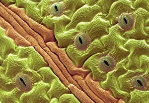
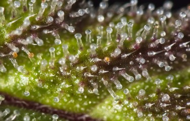
Scanning Eletron Microscopy photograph of the leaf surface of Solanum
By A Mystery Man Writer
Last updated 18 Jun 2024

Download scientific diagram | Scanning Eletron Microscopy photograph of the leaf surface of Solanum granuloso-leprosum Dunal. A – Unicelular tector trichomes; B – tector trichome, note that there are projections at the trichome base; C – tector trichome, note that there is a larger projection/ramification at the trichome base; D – tector trichome, note that there are two larger projection/ramification at the trichome base; E – tector trichome, note that there are three larger projection/ramification at the trichome base; F – tector trichome, note that there are four larger projection/ramification at the trichome base; G – tector trichome, note that there are five larger projection/ramification at the trichome base; H – tector trichome, note that there are six larger projection/ramification at the trichome base; I – tector trichome, note that there are eight larger projection/ramification at the trichome base; J – another angle from the six ramification tector trichome; and K – multicelular and multisseriated tector trichome, note the thick secondary cell wall. Scale Bars = 20 μm. from publication: Anatomy, histochemistry and micromorphology of leaves of Solanum granuloso-leprosum Dunal | In the present work the anatomical, histochemical and micromorphological features of S. granuloso-leprosum leaves were approached in order to evaluate its characteristics associated with its pioneer role. Glandular and non-glandular trichomes were observed on both epidermal | Micromorphology, Solanum and Plant Anatomy | ResearchGate, the professional network for scientists.

A scanning electron microscopy-based screen of leaves of Solanum pennellii (ac. LA716) x Solanum lycopersicum (cv. M82) introgression lines provides a resource for identification of loci involved in epidermal development in tomato
Transpiration Collection of Photo Prints and Gifts
Scanning electron microscopic (SEM) images captured at 60×

SEM micrographs showing surface structure of chemical fixed leaves and

Topography-Driven Shape, Spread, and Retention of Leaf Surface Water Impacts Microbial Dispersion and Activity in the Phyllosphere

Tomato stem, SEM - Stock Image - C021/7479 - Science Photo Library

Scanning electron microscopic images of the lower leaf epidermis
Scanning electron microscope (SEM) images of the foliar trichomes of
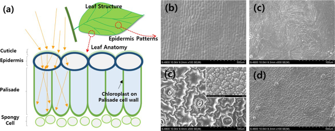
Omnidirectional Light Capture by Solar Cells Mimicking the Structures of the Epidermal cells of Leaves
Recommended for you
-
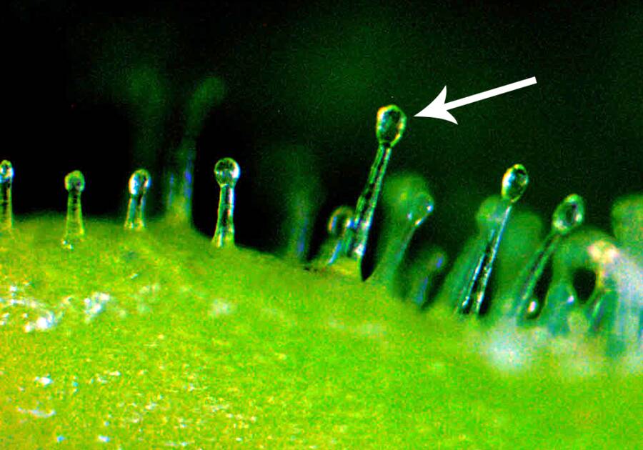 Trichomes18 Jun 2024
 -
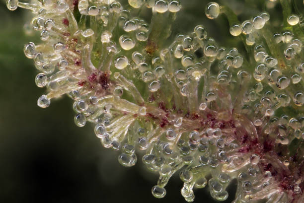 Cannabis Trichomes Stock Photo - Download Image Now - Plant Trichome, Cannabis Plant, Macrophotography - iStock18 Jun 2024
 -
 What are Cannabis Trichomes? An Overview + Growing Tips18 Jun 2024
 -
 How to take trichome pics? - Grow Equipment - I Love Growing Marijuana Forum18 Jun 2024
 -
 Trichome Color Changes Tell You When to Harvest Your Cannabis Buds?18 Jun 2024
 -
 File:Müürlooga (Arabidopsis thaliana) lehekarv (trihhoom) 311 0804.JPG - Wikipedia18 Jun 2024
 -
 SmartPhone Trichome Scope - Easiest way to check on Trichome Maturity18 Jun 2024
 -
 Trichome microscope hack for crystal clear pics with 100% stillness! - Harvesting - I Love Growing Marijuana Forum18 Jun 2024
 -
 Trichome distribution on first leaves of Col and Ler plants. (A) Col.18 Jun 2024
 -
 TOMLOV Wireless Digital WiFi Microscope 50X-1000X Portable USB18 Jun 2024
You may also like
-
 Follow The Yellow Brick Home - Early Spring Styled Tray with18 Jun 2024
 -
 Mini Coloring Book Pocket Size Adult Coloring Hand Drawn18 Jun 2024
 -
 wholesale aircraft grade 1mm 5mm basswood18 Jun 2024
 -
 Airbrush Supplies18 Jun 2024
 -
 Convertible Top Care by RaggTopp – Ask a Pro Blog18 Jun 2024
 -
 Report: Cameo Business Breakdown & Founding Story18 Jun 2024
 -
 Liquitex Professional Acrylic Ink 30ml Yellow Orange Azo18 Jun 2024
 -
🔥FREE SHIPPING🔥Electric Drill Plate Cutter, Universal Metal Nibbler Drill Attachment with Adapter - Drills, Facebook Marketplace18 Jun 2024
-
 Bamboo 16 Inch (40 cm) Circular18 Jun 2024
 -
 KOKNIT Crochet Hooks Set with Storage Case - 9 Soft Grip Ergonomic Crochet Needles,12 Coloured Aluminum Yarn Crocheting Crochet Hooks,Crochet Kit18 Jun 2024
